


INTRODUCTION
Anyone who has spent time studying a drop of blood under a darkfield microscope will have observed a number of different morphologies developing in real-time. As time passes, these morphologies can become more and more complex. The more symptoms the patient has, and particularly with chronic, degenerative diseases, the more complex the “organisms” observed.This phenomenon was observed by leading microscopists such as Antoine Bechamp, Gunther Enderlein, Royal Raymond Rife, Wilhelm Reich, Gaston Naessans, as well as by present-day microscopists. All have seen small particles moving vigorously in body fluids, which were named “protits”, “bions” or “somatids.” Many believed that these moved by Brownian motion, and some scientists believed that they contain genetic material that gives them the ability to reproduce and therefore develop into higher forms of complex morphologies. This theory remains to be proven.
Anyone who has spent time studying a drop of blood under a darkfield microscope will have observed a number of different morphologies developing in real-time. As time passes, these morphologies can become more and more complex. The more symptoms the patient has, and particularly with chronic, degenerative diseases, the more complex the “organisms” observed.This phenomenon was observed by leading microscopists such as Antoine Bechamp, Gunther Enderlein, Royal Raymond Rife, Wilhelm Reich, Gaston Naessans, as well as by present-day microscopists. All have seen small particles moving vigorously in body fluids, which were named “protits”, “bions” or “somatids.” Many believed that these moved by Brownian motion, and some scientists believed that they contain genetic material that gives them the ability to reproduce and therefore develop into higher forms of complex morphologies. This theory remains to be proven.
There has however been research to try to determine the DNA sequences for the complete Cyclogeny as described by Enderlein. Apparently, this research has shown that there is no genetic material in these protits or somatids.
UNANSWERED QUESTIONS
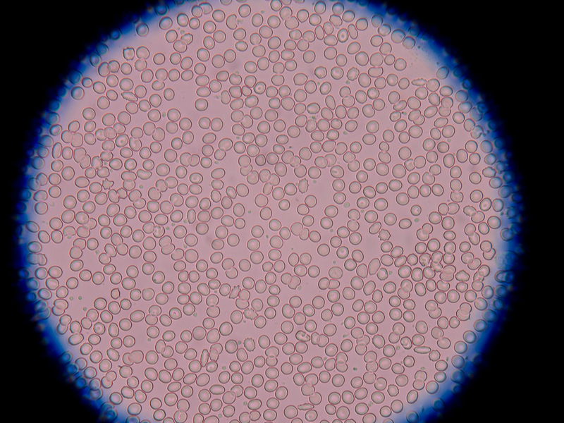
So given that there is no genetic material for these morphologies, then just what is it that we are undeniably viewing in the polymorphic progressions that occur in live blood pictures? How are these complex morphologies actually produced (see images below for examples)? Certainly there have been researchers that have seen realbacteria undergo pleomorphic changes, such as Royal R. Rife who stated that there are a total of 10 different germs and that these change according to the medium that they live in. The pleomorphic development of E. coli,for example, would be:
E. coli to salmonella typhi to mycobacterium tuberculosum to yeast forms to the bacterium X (BX) and finally the bacterium Y (BY).
These are real organisms in the sense that they have genetic structures that have been identified by geneticists. However, the various forms that are seen under darkfield such as ascits, chondroits, diokothecits, filum, mych, protits, synascits, thecits and others have not been identified as having any genetic material. The question stated above regarding how these complex morphologies are produced refers to these “Enderlein” morphologies, not thereal bacteria that were observed by Rife undergoing pleomorphic changes.
A POSSIBLE EXPLANATION OF PLEOMORPHIC CHANGES
During an interesting Live Blood Analysis workshop in London recently, a possible explanation for this “grouping of particles” phenomenon was postulated by one of the authors. It is worthwhile expounding on this concept, as it is possible to explain the flocculation of particles by the physics of ZETA-POTENTIAL.
WHAT IS ZETA-POTENTIAL The zeta potential is a measure of the magnitude of the repulsion or attraction between particles. Simply put, Zeta Potential is a measurement of the charge on the surface of blood cells, platelets, proteins and debris in the plasma. It is the force responsible for particles in blood repelling one another or clumping together.
Around all negatively charged particles there is a charge, which will attract positive ions. The tightly bound positive ion surrounding the particle is known as the stern layer. Ions further away from the particle form the diffuse layer. Somewhere within the diffuse layer is a notional boundary referred to as the Hydrodynamic Plane of Shear.
The zeta-potential is the electrical potential at the hydrodynamic plane of shear. It depends not only upon the particle surface, but also on the dispersant and can be affected by small changes in pH or the ionic strength of the medium. Particles react to the magnitude of the zeta-potential not to their surface charge. Charge interactions between particles probably play a role in all dispersion mechanisms.
HOW IS THIS SIGNIFICANT TO LIVE BLOOD ANALYSIS?
It may well be that the zeta-potential of the blood medium that we are examining under darkfield will differ from patient to patient depending on the conductivity and pH of the medium. The healthier the patient’s blood, the more it will stay within an optimum zeta potential and hence there will be little flocculation of particles. The more the zeta potential moves away from optimum parameters, the more flocculation there will be, leading the microscopist to see the high-valance morphologies seen in the Enderlein Cyclogeny. Let’s look at the logistics of these physical principles a little more carefully.
Blood is intended to be in a dispersed state that is just on the verge of beginning to aggregate. This is required for an effective clotting mechanism, so if we cut ourselves we don’t bleed to death. The clotting cascade is triggered when platelets, activated by damage, release positively charged (cationic) calcium ions together with a series of clotting factors and enzymes. The result is insoluble fibrin threads that form a blood clot. With blood naturally poised at this point, clearly anything we eat, drink or do that pushes it even slightly further in that direction will have a major impact on the blood picture.
Agglutination is influenced by a variety of factors but anything that donates or steals electrons affects Zeta Potential by altering the degree of negative charge on the surface of RBCs and other constituents of plasma. Anything that reduces this negative charge will increase the stickiness of blood, and vice versa.
THE DYNAMICS OF ZETA POTENTIALS
If all the particles have a large zeta-potential (either +ve or –ve) they will repel each other and there is DISPERSION STABILITY. If the particles have low zeta-potentials, there is no force to prevent the particles coming together and there is DISPERSION INSTABILITY, causing particles to flocculate, as observed under the darkfield microscope.In general, the more in the optimum range of zeta potential, the more stable the particle dispersion is likely to be. The dividing line between an aqueous particle dispersion being stable and instability is considered to be +30mV and –30mV. So if all particles have a zeta-potential that is more positive than +30mV or more negative than –30mV, the dispersion should remain stable. The closer the zeta-potentials get to 0mV, the more likely we are to see flocculation or sticking together of particles.
If all the particles have a large zeta-potential (either +ve or –ve) they will repel each other and there is DISPERSION STABILITY. If the particles have low zeta-potentials, there is no force to prevent the particles coming together and there is DISPERSION INSTABILITY, causing particles to flocculate, as observed under the darkfield microscope.In general, the more in the optimum range of zeta potential, the more stable the particle dispersion is likely to be. The dividing line between an aqueous particle dispersion being stable and instability is considered to be +30mV and –30mV. So if all particles have a zeta-potential that is more positive than +30mV or more negative than –30mV, the dispersion should remain stable. The closer the zeta-potentials get to 0mV, the more likely we are to see flocculation or sticking together of particles.
The zeta-potential of particle dispersion can be affected by:
1. Changes in the pH of the sample – even very small changes. In general, the zeta-potential is positive at high pH and negative at low pH (Fig 1). Any pH changes of between 4 – 7.5 will cause flocculation or sticking together of particles. The most serious flocculation would occur at a pH of around 5.5 – this is the isolectric point (see Fig 2). Any pH above 7.5 (most healthy body tissues should remain in this range) will lead to stability of particles and no flocculation.

Fig 2 The Isoelectric Point – 5.5 = serious flocculation
2. The conductivity of the medium (concentration and whether there are minerals present or not, as well as the presence of divalent or trivalent cations).
3. The concentration of a particular additive in the sample, such as xenobiotics, viruses, bacteria, parasites that are part of most chronic diseases.
Zeta-potentials can be measured with a Zetasizer Nano instrument using the Laser Doppler Electrophoresis technique and the patented technique of Mixed Mode Measurement Phase Analysis Light Scattering (M3-PALS). Particles move with a characteristic velocity, which is dependent on the field strength, the dielectric constant of the medium and the zeta-potential.
CONCLUSIONS
The phenomenon of electrophoresis – the movement of a charged particle relative to the liquid it is suspended in under the influence of an applied electric field – is basic physics. The magnitude of the electrostatic interactions between particles can be determined by measuring the zeta-potential of the particle dispersion.
Enderlein was correct in predicting that valence intensification depends on the prevailing pH of the blood or tissue, but believed that the various “bacteria” observed under darkfield would reproduce either asexually by binary fission or budding (auxanogeny) or sexually after preceding nuclear fusion (Probaenogeny). This does, of course, assume that there is genetic material in the protits, which has yet to be proven.
It is feasible to postulate, given that no genetic material has been identified to date to explain this upward mobility by reproduction, that the complex morphologies observed using the darkfield microscope are created by changes in zeta-potential resulting from different levels of pH in the tissues and plasma, as well as the conductivity of the blood. Xenobiotics, bacteria, viruses, parasites, etc. can affect this, which cause changes in the dielectric constant and field strength.
The possibility postulated here is that the healthier the person, the more alkaline is their tissue and blood; hence there is particle stability with no flocculation and no upward movement of the endobiont. The converse indication is that the more unhealthy the person, the more acidic is their tissue and blood, ensuing in a lower observed zeta-potential, with flocculation and the presence of complex morphologies with high valencies in blood samples.
The phenomenon of the upward movement of the endobiont in the cyclogeny, causing high valences and more and more complex morphologies should be further examined based upon these simple physical phenomena described in this brief article. If and when genetic material is identified in the endobiont, then maybe this will open other avenues of research.
WHAT ABOUT THE SANUM REMEDIES?
The other question that needs to be answered is how do the Sanum remedies work, that Prof. Enderlein scrupulously created and undoubtedly work effectively with various dis-eases. Could it be that the remedies are shifting the zeta-potentials in some way? Could it be that they are varying the dielectric constants and field strength? Could it be that they are functioning by changing the quantum fields? All these are interesting questions that need more deliberation. There is no disputing that Sanum remedies do work effectively, but the question is how?
WHAT IMPROVES THE BLOOD PICTURE?
A negative charge on particles entering the bloodstream helps to increase dispersion by enhancing the Zeta Potential on blood colloids. In fact, anything that increases, protects or replenishes the negative charge on the membranes will be beneficial to health.
According to Dr Patrick Flanagan, who has studied the health-enhancing properties of Hunza water, Hydrogen is particularly important, as it is the only carrier of electrons in the body. It should come as no surprise to learn that a plentiful supply of negatively charged hydrogen electrons (anions) is found in all organic matter. However, storage, processing and cooking depletes plant and animal tissue of H-.
Conversely, positively charged cations are found in foods with chemical preservatives, artificial flavours and colours, and pesticide residue, or food that has been overcooked or microwaved. These foods lower the Zeta Potential of the gut and subsequently the blood.
FACTORS THAT ENCOURAGE DISPERTION
There are a number of factors that can help to disperse the agglutinated matter in the blood and therefore enhance Zeta Potentials:
• Essential Fatty Acids (especially Omega-3s)
• Organic, fresh, raw food
• Antioxidants
• Vitamins: A, D E, K, C
• Others: Co Q10, alpha-lipoic acid
• Minerals: Magnesium, Zinc, Selenium, Manganese
• Phytochemicals: Carotenoids, Flavonoids, Proanthocyanadins, Catechins
• Spring Water
• Energy Medicine
• Ionisers
• Maintaining correct pH
• Balanced electrolytes
• Quality air – clean, fresh, sea
FACTORS THAT ENCOURAGE AGGLUTINATION:
Conversely, there are a number of factors that can encourage agglutination such as:
• Insufficient hydration
• Lack of antioxidants
• Excess free radical activity
• Stress (physical or mental)
• Toxins (from air, food, water, environment, drugs, alcohol, dentistry, medical intervention)
• Electromagnetic Radiation
• Insufficient intake or poor conversion of EFAs (particularly O-3)
• Adverse ratio of Omega-6 to Omega-3 HUFAs (specifically EPA & DHA)
• Trans-fats from damaged / processed foods
• Excess saturated or monounsaturated fatty acids
• Imbalance or lack of electrolytes (esp. magnesium)
• Diet that causes increased acidity in the patient (depends on their MT)
• Allergy (inflammatory response)
• Lectins (proteins found in all grains, legumes, dairy and nightshade family plants)
HOW ESSENTIAL FATTY ACIDS AFFECT ZETA POTENTIAL
Phospholipid enzymes surround blood cells, as well as all other cells and their organelles in the body. They also encapsulate and transport triglycerides and cholesterol in the blood.
Phospholipids carry a negative electrical charge on their surfaces. This is due to the fatty acids incorporated in their molecular structure. The more double bonds there are within the fatty acid components of the membrane, the more fluid the structures are and the stronger their negative charge (Udo Erasmus, 1993, Fats that Heal Fats that Kill).
In effect, this negative charge forms a force-field around any particles in the blood surrounded by phospholipids. The stronger the negative charge, the more they repel one another. If the negative charge is reduced the effect will lessen, allowing the constituents of blood to get closer together and begin to aggregate.
Saturated fats have no double bonds and therefore no charge, whilst the long-chain polyunsaturated fatty acids with the most double bonds (Omega-3s: EPA and DHA) carry the strongest negative charge. This fact illustrates the importance of ensuring an adequate intake of essential fatty acids. They directly influence Zeta Potential and that affects the delicate balance of dispersion versus flocculation in the blood.
There is evidence to indicate that the ideal intake of Omega-6 and Omega-3 EFAs should be in a ratio of 1:1. It is important to understand here that most people have a very high intake of Omega-6 EFAs. They are present in all vegetable oils, grains and animal fats, and the foods made from them, which includes the convenience products that form the bulk of many people’s diets. In comparison the best sources of Omega-3s (flaxseed and oily fish) are not consumed very often.
Furthermore O-6s generally have pro-inflammatory effects, and are used to make eicosanoids and prostaglandins that are involved in clotting processes, whilst O-3s are anti-inflammatory and have a thinning effect on the blood.
To redress the imbalance, many nutritionists recommend flaxseed oil supplements but apparently do not realise that often it is of little benefit. Research and practise indicates that large sectors of the populace are not able to convert ALA to the longer chain EPA and DHA that actually produce the anti-inflammatory effects. This applies especially to the chronically ill and those with a genetic adaptation to a diet high in oily fish (ie those with ancestors from temperate and colder climates), as both groups tend to produce insufficient Desaturase enzymes, vital for the conversion. The process is also inhibited by a diet high in carbohydrate and trans-fatty acids, found in most processed foods.
Therefore, to improve the balance between O-6s and O-3s and affect the blood picture positively, supplements of high-quality, concentrated fish-oil are advised, together with natural, full-spectrum vitamin E to protect the oil from free radical attack.
CAUTION: Anyone on blood thinning / anti-coagulation drugs (e.g. Warfarin, aspirin) must consult their GPs and / or Specialists before taking fish oil or vitamin E as these supplements will affect clotting time.
IN SUMMARY
LIVE BLOOD MICROSCOPY is a tool that allows us to directly view the blood and determine how Zeta Potential and other factors are affecting it. It enables us to visually monitor and evaluate a patient’s response to therapeutic measures. Most importantly, it gives patients a window to see into their body, which they sometimes find shocking but often highly motivating.
Understanding the basic concept of Zeta Potential, and utilising that knowledge in conjunction with the powerful medium of visual medicine afforded by the microscope, will enable medical and complementary practitioners to solve many common health problems relatively quickly and inexpensively.
Always bear in mind that neither drugs nor supplements cure disease. The body repairs itself, given the raw materials to do so. Don’t underestimate the importance of the holistic approach. It addresses the underlying cause of disease, rather than isolated symptoms.
There is so much we don’t know, but we do know that nature supplies us with all we need for healthy bodies. A natural, organic diet, pure drinking water, fresh air, sunlight, quality sleep and limited exposure to processed, fake, synthetic foods and drink and commonplace toxins will go a long way towards improving the blood picture. If you add to that basic prescription nothing more than Omega-3 EFAs and a wide range of antioxidants, many patients’ health issues will recede.
Dr George J Georgiou
Holistic Medicine PractitionerDr Georgiou has been an active clinician for 30 years, and Director Founder of the Da Vinci Holistic Health Centre and the Da Vinci College of Holistic Medicine in Larnaca, Cyprus.


KRILL PLUS – 60 caps
Original price was: €30.45.€28.00Current price is: €28.00.
NATURAL ANTIBIOTIC PACK
Original price was: €76.85.€69.25Current price is: €69.25.0 Days
0 Hours
0 Minutes
0 Seconds

SUPER PARASITE DETOX PACK
Original price was: €111.70.€100.75Current price is: €100.75.0 Days
0 Hours
0 Minutes
0 Seconds
Top News